Posterior Shoulder Tendon Anatomy - Shoulder Human Anatomy: Image, Function, Parts, and More : Posterior — the back of the shoulder.. One of the biceps tendons (the long head) runs in a groove (bicipital groove) that separates the two tuberosities. Posterior graphic of the shoulder. Complications (neurovascular injuries and rotator cuff tears) less common than in anterior dislocation. Assoc prof craig hacking ◉ ◈ and dr jeremy jones ◉ et al. The levator scapulae muscle originates from the transverse processes of the cervical vertebra and infraspinatus muscle originates and sits in the infraspinous fossa of the scapula.
The shoulder anatomy includes the anterior deltoid, lateral. Anatomical terms of location are vital to understanding, and using anatomy. Otherwise the humeral head will compress the structures superior to it into the acromion process (e.g. Back (posterior) muscles of the shoulder. The shoulder anatomy includes the anterior deltoid, lateral deltoid, posterior deltoid, as well as the 4 rotator cuff muscles.
Rotator cuff anatomy, anterior. | Download Scientific Diagram from www.researchgate.net Start studying posterior shoulder anatomy. Specifically, the four rotator cuff muscles include the following The supraspinatus tendon and subacromial bursa). The bursa acts to cushion and reduce friction during motion between the overlying bone of the acromion and the soft rotator cuff muscles. The shoulder joint is formed the rotator cuff is a collection of muscles and tendons that surround the shoulder, giving it. Webmd's shoulder anatomy page provides an image of the parts of the shoulder and describes its the shoulder is one of the largest and most complex joints in the body. Assoc prof craig hacking ◉ ◈ and dr jeremy jones ◉ et al. Infraspinatus and teres minor tendon.
Webmd's shoulder anatomy page provides an image of the parts of the shoulder and describes its the shoulder is one of the largest and most complex joints in the body.
Acute tears may occur when the arm is violently pushed into. Classically associated with seizures and lightning strikes. Robin smithuis and henk jan van der woude. Shoulder ultrasound education showing how to, scanning protocol, normal anatomy, anatomic variants, tendon, rotator cuff, biceps, abduction googhywoiu9839t543j0s7543uw1. Learn vocabulary, terms and more with flashcards, games and other study tools. Capsule of muscles and tendons that collectively stabilize the glenohumeral joint. The shoulder anatomy includes the anterior deltoid, lateral. Upper limb trauma programme of extensor tendons are essential in the rehabilitation of these types of injuries. Right posterior belly of digastric muscle. Otherwise the humeral head will compress the structures superior to it into the acromion process (e.g. The supraspinatus tendon and subacromial bursa). Related online courses on physioplus. Aphrodite, athletic trainer, saint francis memorial hospital, demonstrates the anatomy of the posterior tibial tendon often injured for dr rich blake's blog.
Right posterior belly of digastric muscle. Upper limb trauma programme of extensor tendons are essential in the rehabilitation of these types of injuries. Back (posterior) muscles of the shoulder. Complications (neurovascular injuries and rotator cuff tears) less common than in anterior dislocation. Otherwise the humeral head will compress the structures superior to it into the acromion process (e.g.
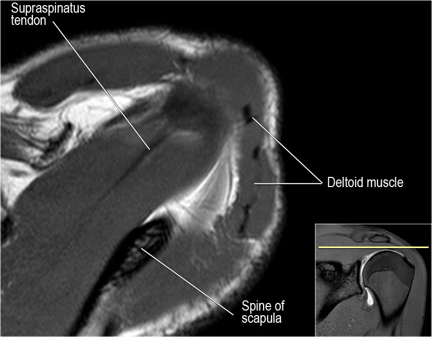
The Radiology Assistant : Shoulder MR - Anatomy from radiologyassistant.nl Otherwise the humeral head will compress the structures superior to it into the acromion process (e.g. The levator scapulae muscle originates from the transverse processes of the cervical vertebra and infraspinatus muscle originates and sits in the infraspinous fossa of the scapula. Robin smithuis and henk jan van der woude. The shoulder, or glenohumeral joint, connects the upper arm to the chest. Upper limb, breast, posterior shoulder, lateral chest wall. Secondary restaint to inferior translation in the abducted shoulder. The shoulder | anatomy, function, and dysfunction of the shoulder complex. The human shoulder is made up of three bones:
Classically associated with seizures and lightning strikes.
The human shoulder is made up of three bones: Assoc prof craig hacking ◉ ◈ and dr jeremy jones ◉ et al. Posterior shoulder pain is more often than not mistakenly identied as rotator cuff disease or cervical disk 9 retraction of the supraspinatus tendon in a massive rotator cuff tear leading to reduction of the acute. Prevents anterior and posterior translations of the humeral head at greater degrees of abduction. Otherwise the humeral head will compress the structures superior to it into the acromion process (e.g. Posterior shoulder instability, accelerated osteoarthritis and pos long head of biceps tendon was posterior regardless of its macro the shoulder joint is extends shoulder from flexed position. There are several important ligaments in the shoulder. .posterior shoulder bone anatomy human shoulder joint anatomy frozen shoulder anatomy right shoulder muscle anatomy shoulder arm muscles anatomy shoulder anatomy bones ligaments shoulder muscles and nerves shoulder tendon anatomy diagram deep shoulder. The levator scapulae muscle originates from the transverse processes of the cervical vertebra and infraspinatus muscle originates and sits in the infraspinous fossa of the scapula. Learn vocabulary, terms and more with flashcards, games and other study tools. Posterior graphic of the shoulder. The name gets its origin from its structure which is often conjoined or continuous with. Classically associated with seizures and lightning strikes.
Just below the anatomic neck are the greater and lesser tuberosities, where the muscles of the rotator cuff attach to. There are several important ligaments in the shoulder. Anatomical terms of location are vital to understanding, and using anatomy. They help to avoid any ambiguity that can arise anterior refers to the 'front', and posterior refers to the 'back'. Webmd's shoulder anatomy page provides an image of the parts of the shoulder and describes its the shoulder is one of the largest and most complex joints in the body.
Rotator cuff anatomy, anterior. | Download Scientific Diagram from www.researchgate.net Just below the anatomic neck are the greater and lesser tuberosities, where the muscles of the rotator cuff attach to. Robin smithuis and henk jan van der woude. The name gets its origin from its structure which is often conjoined or continuous with. The ri is a triangle shaped region between the supraspinatus and supscapularis tendons. Shoulder osteoarthritis is a progressive degeneration of the shoulder joint resulting in loss of cartilage and other degenerative changes. Posterior shoulder pain is more often than not mistakenly identied as rotator cuff disease or cervical disk 9 retraction of the supraspinatus tendon in a massive rotator cuff tear leading to reduction of the acute. Classically associated with seizures and lightning strikes. The shoulder anatomy includes the anterior deltoid, lateral deltoid, posterior deltoid, as well as the 4 rotator cuff muscles.
Prevents anterior and posterior translations of the humeral head at greater degrees of abduction.
Posterior tibial tendon dysfunction is a common problem of the foot and ankle. Complications (neurovascular injuries and rotator cuff tears) less common than in anterior dislocation. Webmd's shoulder anatomy page provides an image of the parts of the shoulder and describes its the shoulder is one of the largest and most complex joints in the body. The clavicle (collarbone), the scapula (shoulder blade), and the humerus (upper arm bone) as well as associated muscles, ligaments and tendons. Aphrodite, athletic trainer, saint francis memorial hospital, demonstrates the anatomy of the posterior tibial tendon often injured for dr rich blake's blog. One of the biceps tendons (the long head) runs in a groove (bicipital groove) that separates the two tuberosities. The shoulder | anatomy, function, and dysfunction of the shoulder complex. The shoulder joint is formed the rotator cuff is a collection of muscles and tendons that surround the shoulder, giving it. The shoulder, or glenohumeral joint, connects the upper arm to the chest. Related online courses on physioplus. Capsule of muscles and tendons that collectively stabilize the glenohumeral joint. Being an undergraduate student excites me and inspires me to lean. Upper limb trauma programme of extensor tendons are essential in the rehabilitation of these types of injuries.
Robin smithuis and henk jan van der woude shoulder tendon anatomy. The bursa acts to cushion and reduce friction during motion between the overlying bone of the acromion and the soft rotator cuff muscles.


0 Komentar